


![Anti-alpha 1 Fetoprotein antibody [EPAFP61] 40μl](https://yunshiji.oss-cn-shenzhen.aliyuncs.com/202407/25/mlko2co1hdp.jpg)
![Anti-alpha 1 Fetoprotein antibody [EPAFP61] 100μl](https://yunshiji.oss-cn-shenzhen.aliyuncs.com/202407/25/i22chgsp01w.jpg)
![Anti-alpha 1 Fetoprotein antibody [EP1017Y] 10μl](https://yunshiji.oss-cn-shenzhen.aliyuncs.com/202407/25/1k5e2gdxdtr.bmp)
![Anti-alpha 1 Fetoprotein antibody [EP1017Y] 40μl](https://yunshiji.oss-cn-shenzhen.aliyuncs.com/202407/25/snhokg5ajpm.bmp)
![Anti-alpha 1 Fetoprotein antibody [EP1017Y] 100μl](https://yunshiji.oss-cn-shenzhen.aliyuncs.com/202407/25/frcrjdmh1pg.bmp)
![Anti-alpha 1 Fetoprotein antibody [EP1016Y] 10μl](https://yunshiji.oss-cn-shenzhen.aliyuncs.com/202407/25/qbwrj5bto3w.gif)
詳細(xì)說明
概述
產(chǎn)品名稱Anti-alpha Actinin抗體[EP2527Y]
描述
兔單克隆抗體[EP2527Y] to alpha Actinin
經(jīng)測試應(yīng)用WB,IP,Flow Cyt,IHC-P
種屬反應(yīng)性
與反應(yīng): Mouse, Rat, Human
免疫原
A synthetic peptide corresponding to residues on the N-terminus of human alpha Actinin
陽性對照
NIH 3T3 lysate, HeLa lysate and C6 lysate
常規(guī)說明
Produced using Abcam’s RabMAb? technology. RabMAb? technology is covered by the following U.S. Patents, No. 5,675,063 and/or 7,429,487.
性能
形式Liquid
存放說明Shipped at 4°C. Upon delivery aliquot and store at -20°C. Avoid freeze / thaw cycles.
存儲溶液pH: 7.40
Preservative: 0.01% Sodium azide
Constituents: 50% Glycerol, 0.05% BSA純度Tissue culture supernatant
克隆單克隆
克隆編號EP2527Y
同種型IgG
研究領(lǐng)域
Developmental Biology
Lineage specification
Mesoderm
Stem Cells
Lineage Markers
Mesoderm
Signal Transduction
Cytoskeleton / ECM
Cytoskeleton
Microfilaments
Actin etc
Actin Binding Proteins
<a javascript:void(0)>Signal Transduction
Cytoskeleton / ECM
Cytoskeleton
Microfilaments
Actin etc
Actin Crosslinking
Anti-alpha Actinin antibody [EP2527Y] 圖像
![Flow Cytometry - Anti-alpha Actinin antibody [EP2527Y] (ab68194)](http://img.lianshimall.com/statics/attachment/goods/pl20160426/abcamMainImgPrimary/detail/ab6/ab681944FC.jpg)
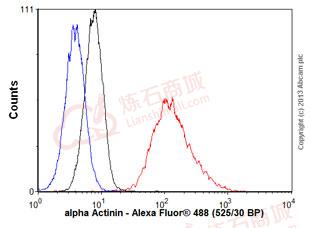
Flow Cytometry - Anti-alpha Actinin antibody [EP2527Y] (ab68194)
Overlay histogram showing HeLa cells stained with ab68194 (red line). The cells were fixed with 80% methanol (5 min) and then permeabilized with 0.1% PBS-Tween for 20 min. The cells were then incubated in 1x PBS / 10% normal goat serum / 0.3M glycine to block non-specific protein-protein interactions followed by the antibody (ab68194, 1/1000 dilution) for 30 min at 22°C. The secondary antibody used was a goat anti-rabbit Alexa Fluor? 488 (IgG; H&L) (ab150077) at 1/2000 dilution for 30 min at 22°C. Isotype control antibody (black line) was rabbit IgG (monoclonal) (0.1μg/1x106 cells) used under the same conditions. Unlabelled sample (blue line) was also used as a control. Acquisition of >5,000 events were collected using a 20mW Argon ion laser (488nm) and 525/30 bandpass filter. This antibody gave a positive signal in HeLa cells fixed with 4% paraformaldehyde (10 min)/permeabilized with 0.1% PBS-Tween for 20 min used under the same conditions.
![Western blot - alpha Actinin antibody [EP2527Y] (ab68194)](http://img.lianshimall.com/statics/attachment/goods/pl20160426/abcamMainImgPrimary/detail/ab6/ab68194wb1.jpg)
Western blot - alpha Actinin antibody [EP2527Y] (ab68194)
All lanes : Anti-alpha Actinin antibody [EP2527Y] (ab68194) at 1/10000 dilution
Lane 1 : NIH 3T3 lysate
Lane 2 : HeLa lysate
Lane 3 : C6 lysate
Lysates/proteins at 10 μg per lane.
Secondary
HRP labelled goat anti-rabbit at 1/2000 dilution
Predicted band size : 103 kDa
Observed band size : 103 kDa
![Immunohistochemistry (Formalin/PFA-fixed paraffin-embedded sections) - Anti-alpha Actinin antibody [EP2527Y] (ab68194)](http://img.lianshimall.com/statics/attachment/goods/pl20160426/abcamMainImgPrimary/detail/ab6/ab68194hcp.jpg)
Immunohistochemistry (Formalin/PFA-fixed paraffin-embedded sections) - Anti-alpha Actinin antibody [EP2527Y] (ab68194) This image is courtesy of an Abreview submitted by Kadir Demircan
ab68194 staining alpha Actinin in rat kidney tissue sections by Immunohistochemistry (IHC-P - paraformaldehyde-fixed, paraffin-embedded sections). Tissue was fixed with formaldehyde and blocked with 30% BSA for 10 minutes at 37°C; antigen retrieval was by heat mediation in EDTA. Samples were incubated with primary antibody (1/100 in PBS) for 16 hours at 4°C. ab80436 EXPOSE Mouse and Rabbit Specific HRP/DAB Detection IHC kit was used.
See Abreview


 粵公網(wǎng)安備44196802000105號